
Home News and Research An insight into how infection can spread
An insight into how infection can spread
Horse A lives in the same field as Horse C but they are separated by electric fencing. Their field is surrounded by agricultural land and gardens, and a bridleway runs down one edge of the field. Both paddocks are thoroughly poo picked every day to two days. Both A and C have been at low burden status since 2012 when ADB first started tapeworm research.
In February 2015, Horse A picked up a tapeworm burden, as determined by an increase in saliva score (Figure 1).
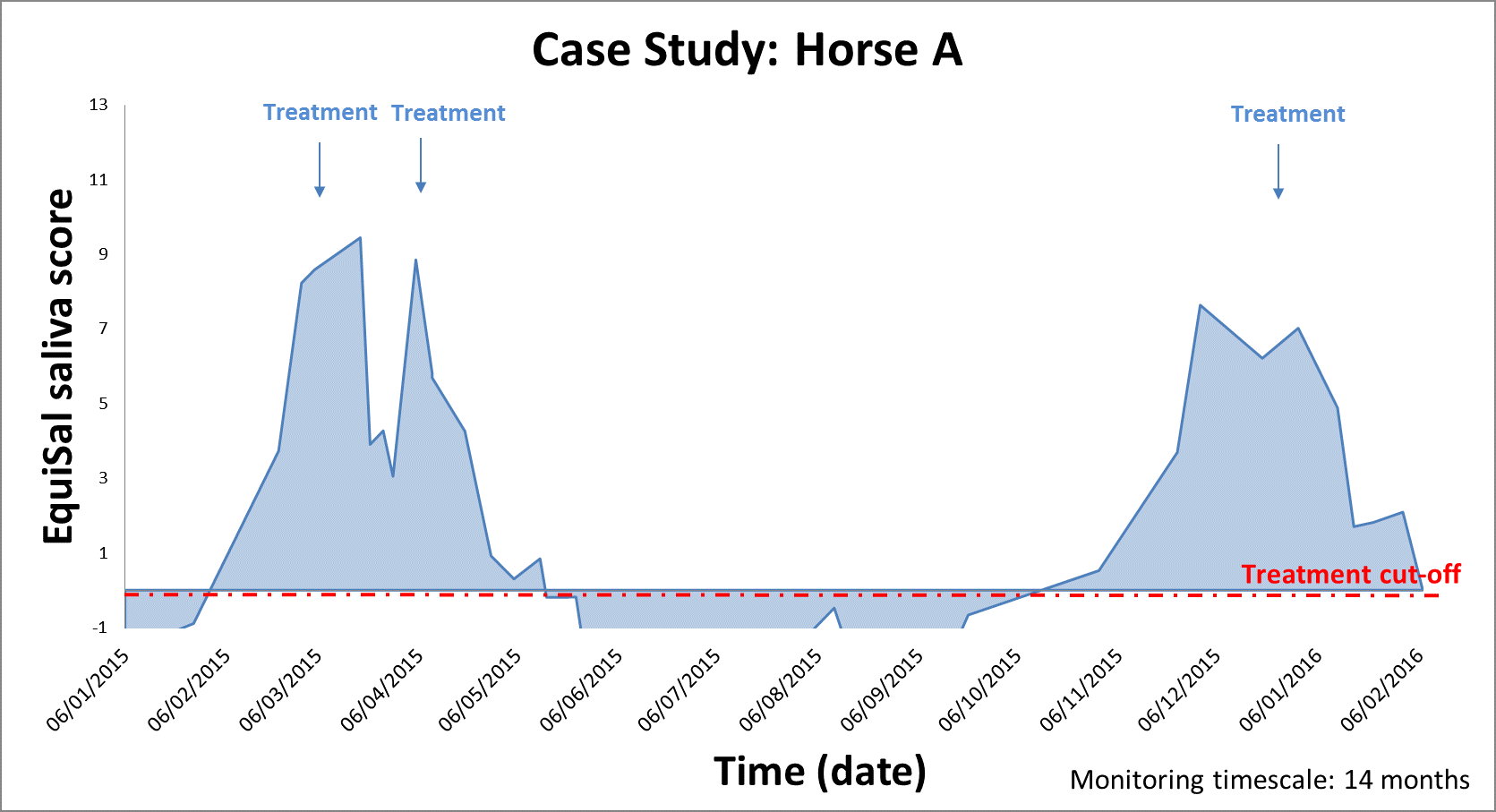
Figure 1. Horse A saliva score monitoring with treatment events
This finding was unexpected as the horses do not leave the paddock and there are no surrounding paddocks that could have infected Horse A. However, it was later found that a horse, passing on the bridleway, had defecated adjacent to the paddock. This may be the first anecdotal evidence that oribatid mites are able to travel from the site of infection, as the mites would have needed to ingest tapeworm eggs from the faeces and then moved to a position where Horse A could have ingested them (Figure 2).

Figure 2. Satellite image detailing the grazing arrangements of Horse A and C from February 2015 to November 2015. Horse A is represented by the brown horse image, while Horse C is represented by the white horse image. The contaminating faeces position is marked with a red star shape.
Horse A’s burden was treated with praziquantel and the saliva score reduced, but then rose again at which point Horse A was given a second treatment. The saliva score reduced to low burden status and remained low for the duration of the summer. However, in November 2015 Horse A’s saliva score increased to moderate/high burden status (Figure 1). Again this was unexpected as the bridleway had been poo picked since the contaminating faeces incident. It is thought that during Horse A’s initial infection, tapeworm eggs would have been released onto the pasture, as illustrated by the red stars in Figure 3.

Figure 3. Satellite image detailing the grazing arrangements of Horse A and C from February 2015 to November 2015. Horse A is represented by the brown horse image, while Horse C is represented by the white horse image. Oribatid mites across the paddock become infected by tapeworm eggs released in the faeces of Horse A, as indicated by the red stars.
Once oribatid mites on the paddock ingested the eggs, the larvae would take at least 2 to 4 months to develop before infecting Horse A again. This is the likely reason that Horse A remained at low burden status throughout the summer before picking up the second burden – as the paddock was previously uncontaminated by tapeworm infections, the larvae required development in the oribatid mite before Horse A could pick up another infection.
During this time, Horse C remained at Low burden status. However, in November 2015, Horse C had his paddock area extended for the winter (Figure 4) and has been grazing part of the paddock normally grazed by Horse A. At the end of January 2016, Horse C was diagnosed with a moderate/high burden. Both horses were treated with praziquantel when they were diagnosed with a burden and have now reduced to low status.

Figure 4. Satellite image detailing the grazing arrangements of Horse A and C from November 2015 to present (Feb 2016). Horse A is represented by the brown horse image, while Horse C is represented by the white horse image. Extension of the paddock for Horse C results in exposure to potentially contaminated pasture, as indicated by the red stars.
What will happen next!